What is the Osteopathic Atlas treatment?
The atlas treatment involves the use of two systems. Firstly, having use of a motion laboratory a biomechanical scan of the body is performed. This scan allows us to create a 3D image of the spine, pelvis, legs, feet and their function. The treatment part of the therapy involves a device that creates a pulse and vibration working through the muscles at the top of the neck. As it works through the soft tissues it is safe for all ages and there are no spinal manipulations involved. The atlas treatment is not invasive. Once the treatment is over the biomechanical scan is performed again. This is used to compare the differences and to show the patient the changes that have ocurred after treatment. The treatment we provide on the atlas is a corrective fix, not a quick fix.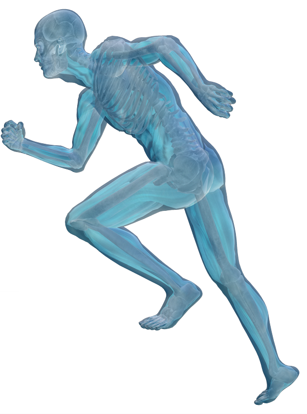
What are the symptoms of an Atlas/neck problem?
An issue with the function of the atlas can have many different symptoms. Some of the symptoms associated with it may be causing pain in the low back for example. Basically our bodies are designed to keep our eyes level. If the atlas is moving incorrectly or poorly within its joints articular surface it causes a side-bending and rotation of the head. In turn, the body compensates by side-bending in the opposite direction, to keep the eyes level. It is this compensation that creates strain and pain patterns, whether they be headaches, neck, back, pelvis or hip pain etc. Most people are unaware that they have an atlas issue, but may be aware that one shoulder appears lower than the other. They may have been told they have one leg shorter than the other or have dropped arches. All these findings can indicate an atlas issue. On an x-ray or MRI some consultants may look at the position of the atlas. However most of the time these scans look at structure rather than function. We diagnose an atlas misalignment via clinical findings.
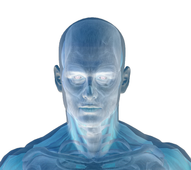
Atlas treatment
What causes the Atlas issue?
The atlas may become an issue in some people following their birth, especially if it was quite difficult or if aids had to be used. Otherwise road traffic accidents (whiplash), traumas to the head neck or shoulders, bad falls or from some reports by patients they felt undergoing forceful dental work can cause the misalignment. Symptoms may develop immediately, such as headaches, neck, back, pelvic pain or may develop over a period of time following the onset.
Where can I get the atlas treatment?
W are the only clinic providing this type of treatment in Ireland. We are the only center in Europe offering the biomechanical assessment as part of the therapy. Other similar treatments on the market include Atlas Orthogonal and Atlas Profilax. We feel that theses therapies do not perform sufficient analysis of the effect of the atlas misalignment and therefore cannot treat it correctly. If you would like to assess whether you have an Atlas problem but would prefer to get your own Doctor, Physiotherapist, Chiropractor or Osteopath to check it? We can instruct your practitioner over the phone on how to analyse it or simply ask them to palpate the Transverse Processes (T.P) of the Atlas. The T.P’s lie inferior to the Mastoid bone, they are looking to assess the position of the atlas in relation to the mastoid. If it is misaligned the T.P will be deeper in on one side in comparison to the other.